What the surgical microscope shows, and what it cannot
Silicon Valley Endodontics and Microsurgery editorial team
Clinical review pending: this page was rewritten and is not covered by any earlier clinical review. Please ask Dr. Kung about your own situation.
An operating microscope magnifies and lights the parts of a tooth we can see directly, such as the pulp chamber floor and canal openings. It helps us look more carefully. It does not let us see through roots, it does not catch every crack, and it cannot guarantee a result. This page explains what the microscope does, what it does not do, and where our information comes from.
What is the microscope for?
The microscope enlarges the treatment area and shines light along the same path we look through. That makes it easier to see small details on surfaces we can reach, such as the pulp chamber floor, canal openings (orifices), and, during surgery, the cut root end. Better visibility can help with finding canal openings and checking our work. It is one tool among many, not a guarantee.
What can the microscope not see?
The microscope only shows what is in its line of sight. It cannot see through a root, around a curve, or inside bone. Some cracks are hidden below the surface or run where no instrument or light can reach, so a crack can remain undetected even with careful magnification. Treatment outcomes depend on many factors, including the tooth, its history, and healing, and are not guaranteed.
What does the 200 to 300 mm focus range mean?
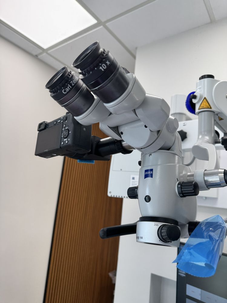
Our clinic has a ZEISS microscope marked Varioskop 100. According to the manufacturer, the Varioskop 100 lets the doctor change the working distance, meaning the space between the microscope lens and the area being viewed, from about 200 to 300 mm without moving the microscope. This is a manufacturer specification about comfortable focusing distance. It is not how deep the microscope can see into a tooth.
What kind of light does it use?
The manufacturer describes LED illumination as an available configuration for this microscope family. We have not confirmed which light source is installed on each of our microscopes, so we do not claim a specific type here. Whatever the source, the light reaches only the surfaces in view.
Can you take pictures through the microscope?
The microscope shown on this page has a camera attached, which can capture images of what is visible through the microscope. Images may be used to explain findings to you or to share with your referring dentist when appropriate. The manufacturer offers optional recording and streaming features, but this page does not describe which options, resolutions, or mounting configuration are installed in our rooms. Our clinic has more than one microscope, and not every room uses the same model.
See examples in our case gallery, or compare approaches in microscope vs. no microscope.
How does the microscope relate to CBCT?
They answer different questions. The microscope uses visible light to view surfaces we can see directly. CBCT is a 3D X-ray scan that can show structures inside bone and roots. CBCT is used for selected cases only, when its information is expected to change decisions. The microscope does not replace X-ray imaging, and CBCT does not replace looking directly.
Learn more about CBCT 3D imaging and our other technology.
What does the research say?
Published reviews describe the operating microscope as part of modern endodontic microsurgery, which combines magnification, lighting, small instruments, ultrasonic root-end preparation, and modern filling materials. A 2010 meta-analysis by Setzer and colleagues compared microsurgical and older surgical techniques as whole approaches. Because these elements are used together, studies do not show that the microscope alone guarantees success. The ZEISS sources listed below are commercial manufacturer descriptions, not independent clinical evidence, and not an endorsement of our clinic.
Common questions
- Does the microscope mean my treatment will succeed?
- No. It improves what we can see on accessible surfaces, but success depends on many factors and cannot be guaranteed.
- Can the microscope find every crack?
- No. Cracks below the surface or out of the line of sight can remain undetected.
- Is the 200 to 300 mm range how deep it sees?
- No. It is the manufacturer's stated working distance between the lens and the treatment area.
- Will I need a CBCT scan?
- Only in selected cases. Dr. Kung will explain if a 3D scan is recommended and why.
Sources
- Independent clinical overviewCarr GB, Murgel CAF. The use of the operating microscope in endodontics. Dent Clin North Am. 2010
- Independent meta-analysisSetzer FC et al. Outcome of endodontic surgery: a meta-analysis. J Endod. 2010
- Manufacturer description (commercial, not clinical evidence)ZEISS OPMI pico product page (current online version mentions optional 4K)
- Manufacturer description (commercial, not clinical evidence)ZEISS OPMI pico international brochure, 2023 edition: pp. 3-4 optics, LED, 200-300 mm focus; pp. 6-7 optional full HD recording/streaming and floor, ceiling or wall configurations
Questions about your tooth?
We are happy to explain what we see, what we cannot see, and what your options are.