What a saved tooth actually looks like
Before-and-after radiographs from real patients treated by Dr. Kung — including the long-term recall films that show infection replaced by healthy, regrown bone. These cases are drawn from the documented portfolio Dr. Kung prepared for the American Board of Endodontics certification process.
Case 01 · Root canal treatment
Lower first molar with four canals — healed at 16 months
This patient's lower molar had deep decay that reached the nerve, causing infection and bone loss around the root tips. Instead of extraction, the tooth was treated with microscope-assisted root canal therapy. Lower first molars usually have three canals — under the microscope, this one revealed four. All four were cleaned, disinfected, and sealed in full. The 16-month recall film shows the result that matters: the infected dark areas are gone and solid bone has grown back.
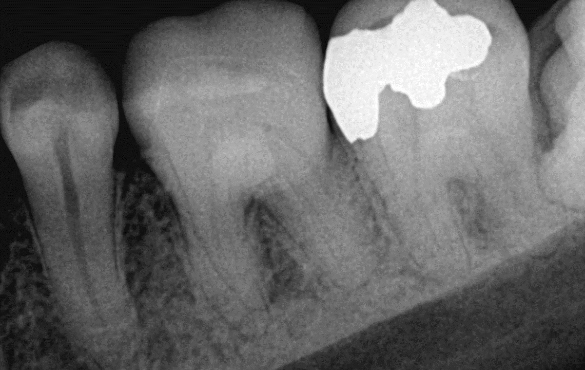 Before
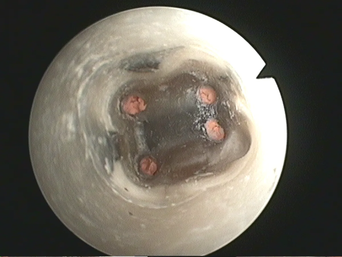 Under the microscope
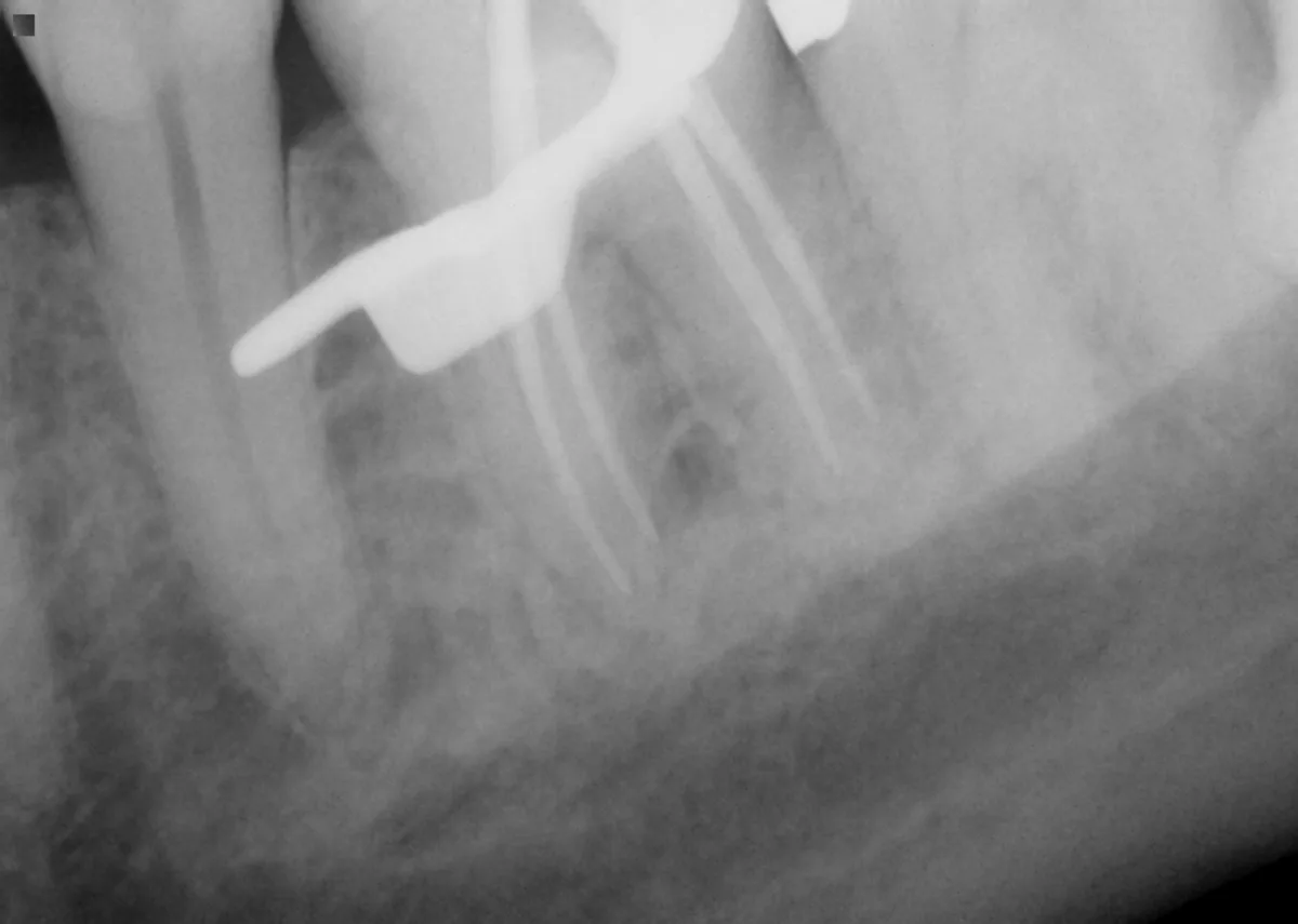 Verification
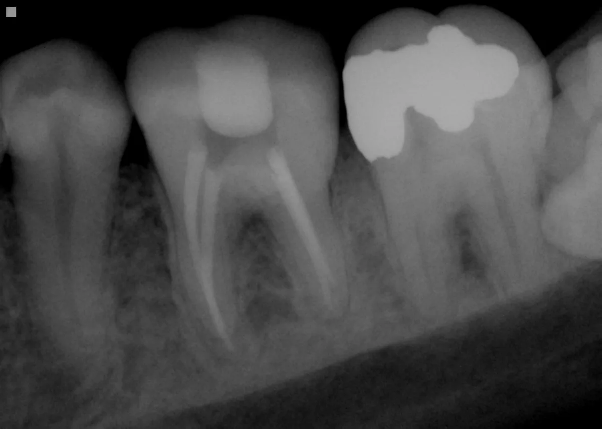 After
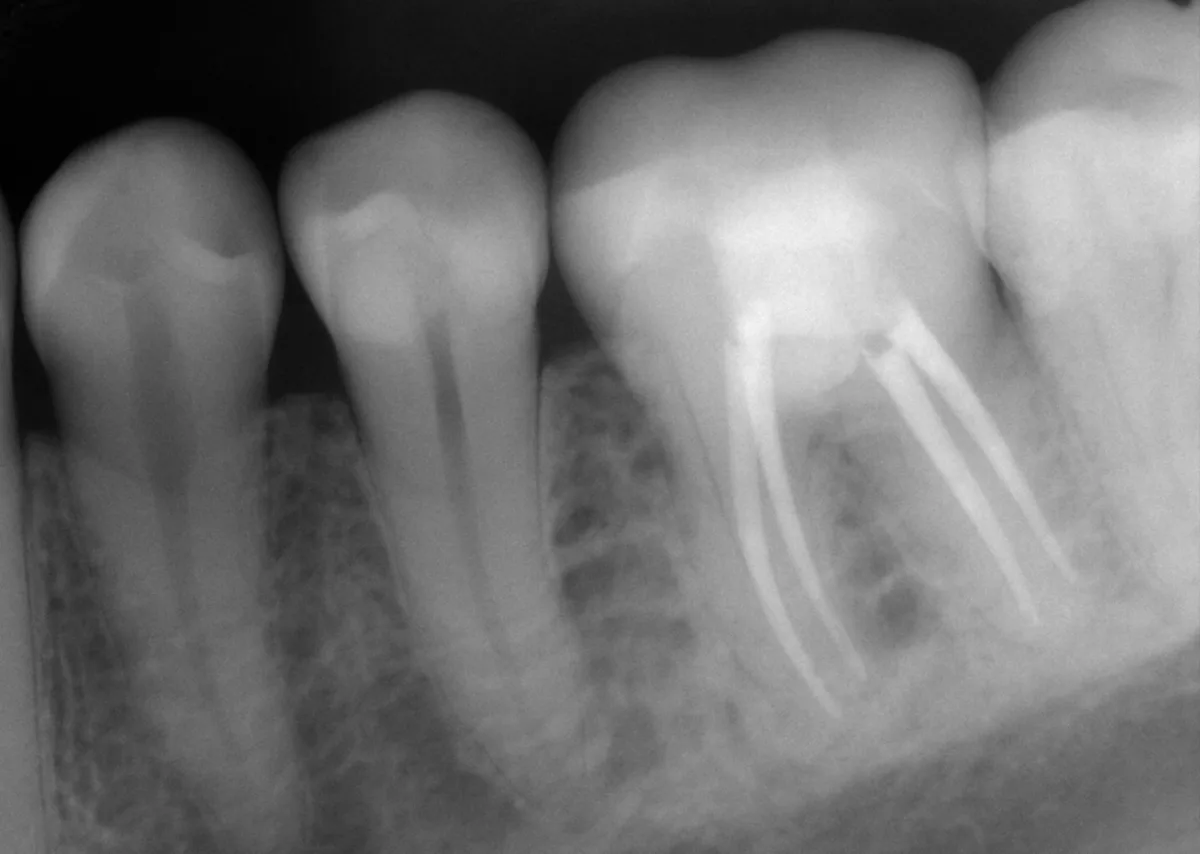 16-month recall
Shown with patient consent; identifying information removed. Individual results vary — no gallery can predict the outcome of your specific tooth. Success depends on the tooth's anatomy, the extent of infection, and timely final restoration.
The fourth canal is the point
Missed canals are one of the leading reasons root canals fail. The microscope photo above shows why specialist treatment is different: that fourth canal is nearly invisible to the naked eye. Every case at our office is treated under a surgical operating microscope, start to finish.
Questions about these cases
Are these real patient cases?
Yes. Every case shown here is a real patient treated by Dr. Jason Kung, shown with the patient's consent and with all identifying information removed. These radiographs are from the case portfolio Dr. Kung prepared for the American Board of Endodontics certification process, which requires fully documented cases with long-term follow-up.
Will my result look like this?
No two teeth are the same, and outcomes vary with the tooth's anatomy, how long the infection has been present, and how the tooth is restored afterward. Published success rates for specialist root canal treatment are high — but a gallery shows representative cases, not a guarantee. At your consultation Dr. Kung will give you an honest assessment of your specific tooth.
What am I looking at in these X-rays?
In the before images, the dark area around the root tip is bone the infection has dissolved. In the after images, the bright white lines inside the roots are the filled, sealed canals. In the recall images taken months later, the dark area has filled back in with healthy bone — that regrowth is what 'healed' looks like on an X-ray.
Wondering if your tooth can be saved?
Most teeth that look hopeless on an X-ray can be treated and kept. Send us your X-ray for a free case assessment, or call — same-day emergency visits available.